3D microscopy workbench, visualization and analysis

MADI3D is a desktop application for organizing, annotating and analyzing raw microscopy data, OBJ meshes and SWC neuronal reconstructions.

Render different data types together, explore multichannel volumes and 4D time series, and inspect spatial relationships with stereoscopic rendering. Stitch acquisitions, register volumes, segment structures, generate Color-Depth MIPs and compare light and electron microscopy data in the same scene.

For Windows, macOS and Ubuntu.

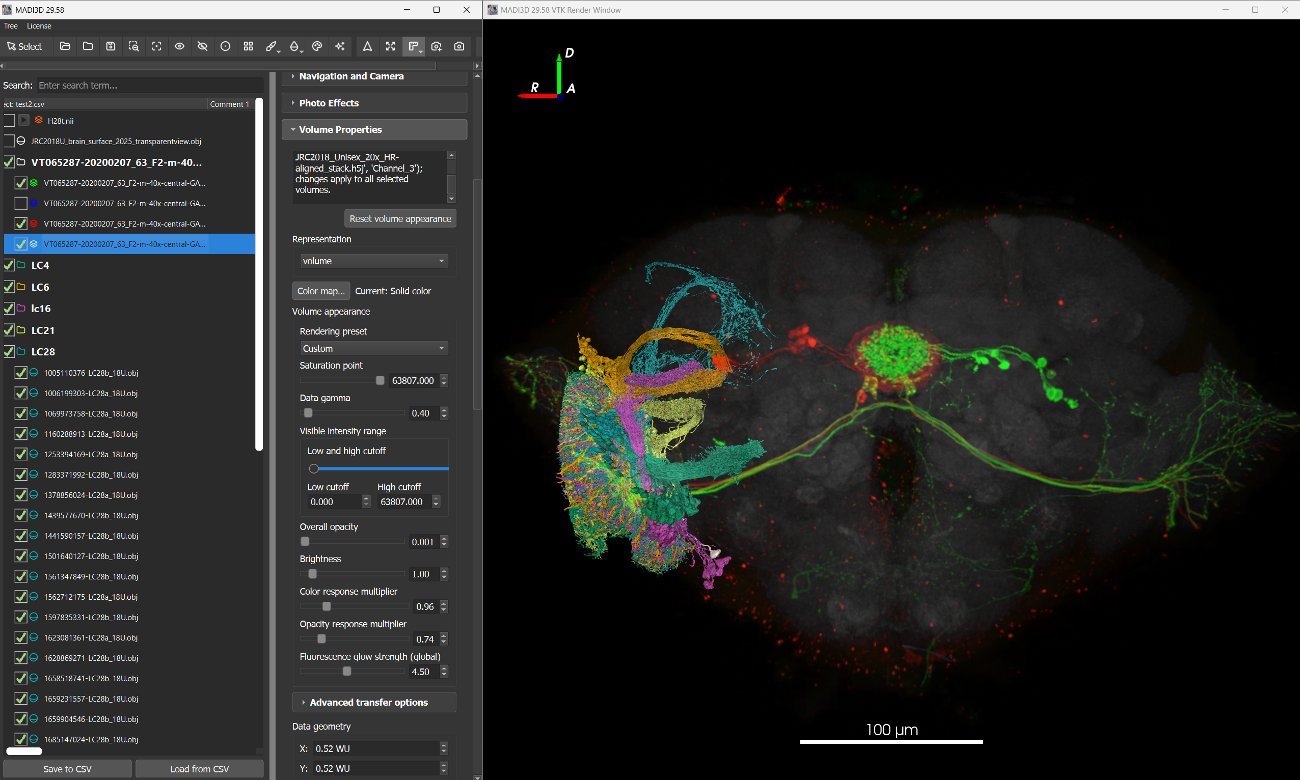
Microscopy volumes and coloured neuronal reconstructions displayed together in MADI3D
Microscopy volumes and neuronal reconstructions in a shared 3D scene. Select any screenshot to view it at full size.

Organize, annotate and explore your data

Keep related datasets and your notes together in a saved project. Geometry, transforms and relationships between datasets remain part of that project.

Project organization

Arrange objects into groups and subgroups, reorder them, and work with individual objects or multiple selections. Organize your project by experiment, specimen or analysis task, then save it to continue your work later. Build storyboards with camera, visibility, object-motion and clipping actions; trim recorded takes and preview the result.

Free-form comment fields

Record experimental details, anatomical labels, observations and review notes in your own words. Multiple comment fields keep these annotations alongside your data, and your notes are saved with the project across sessions.

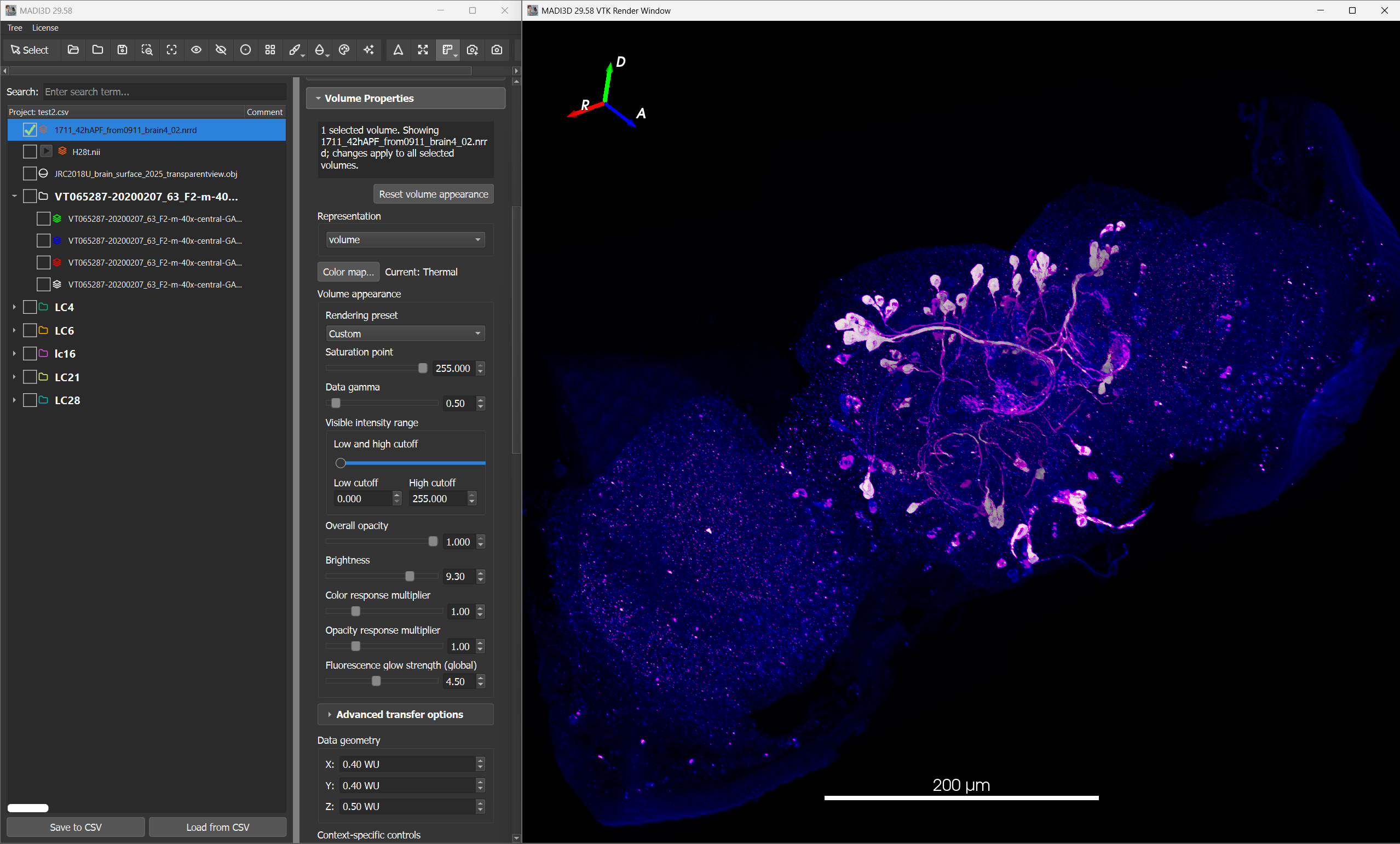
Fluorescence volume rendering and volume controls in MADI3D

Multimodal rendering in 3D and 4D

View raw microscopy volumes, OBJ meshes and SWC neuronal reconstructions together in a shared scene. Explore multichannel data and 4D time series (3D plus time). Use stereoscopic rendering to examine depth and spatial relationships while keeping the source data unchanged.

Import read-only ZEISS CZI grayscale volumes and Nikon ND2 acquisitions with explicit scene or position and channel selection. PLY meshes are also supported; unsafe or ambiguous microscopy layouts are rejected.
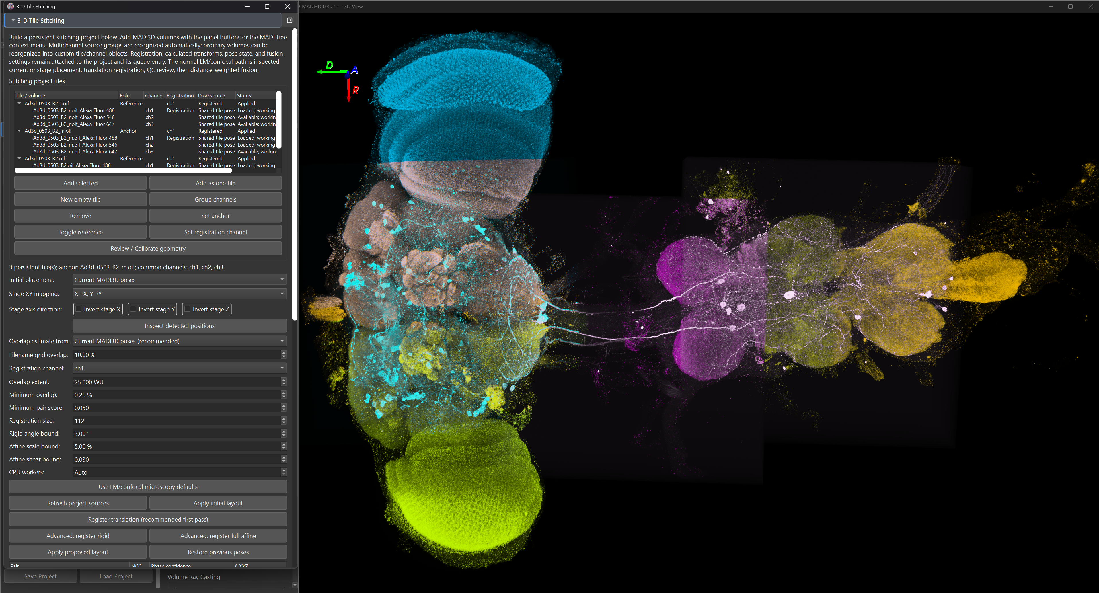
Tiled 3D microscopy acquisitions in the MADI3D stitching workflow

Microscopy stitching

Discover overlapping 3D acquisitions automatically, review their placement and combine tiles into a larger volume. Successful alignment updates the scene; apply and restore controls support review before fusion. Automatic-placement retries retain the selected registration model, and solved geometry can be applied across related channels.

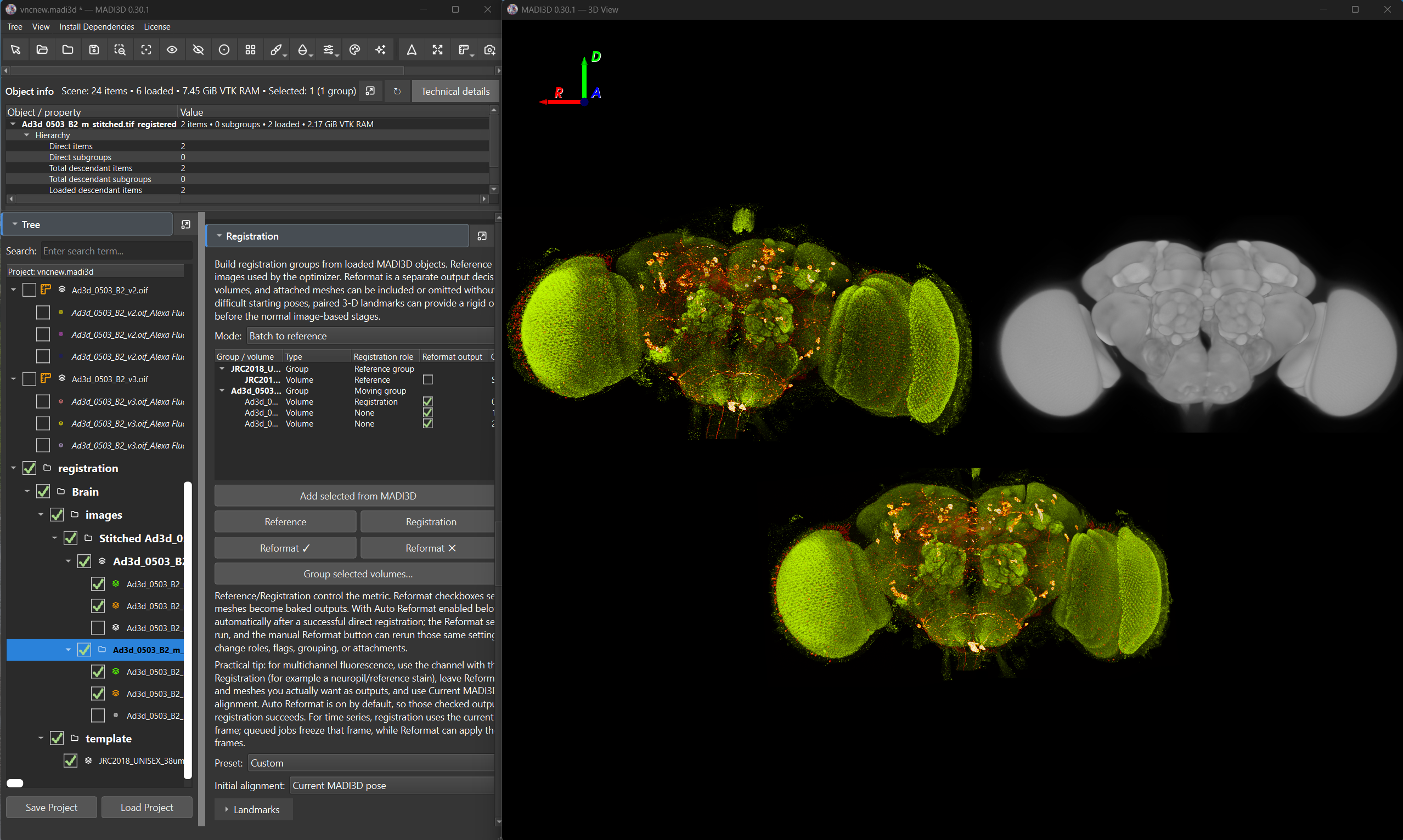
Volume registration using CMTK in MADI3D

Volume registration

Align volumes to a reference anatomy with integrated CMTK registration. Review measured quality checks and configurable thresholds alongside the reference data, and retain transforms for reformatting related channels. Reformatted outputs preserve scene groups, channel labels and scientific geometry.

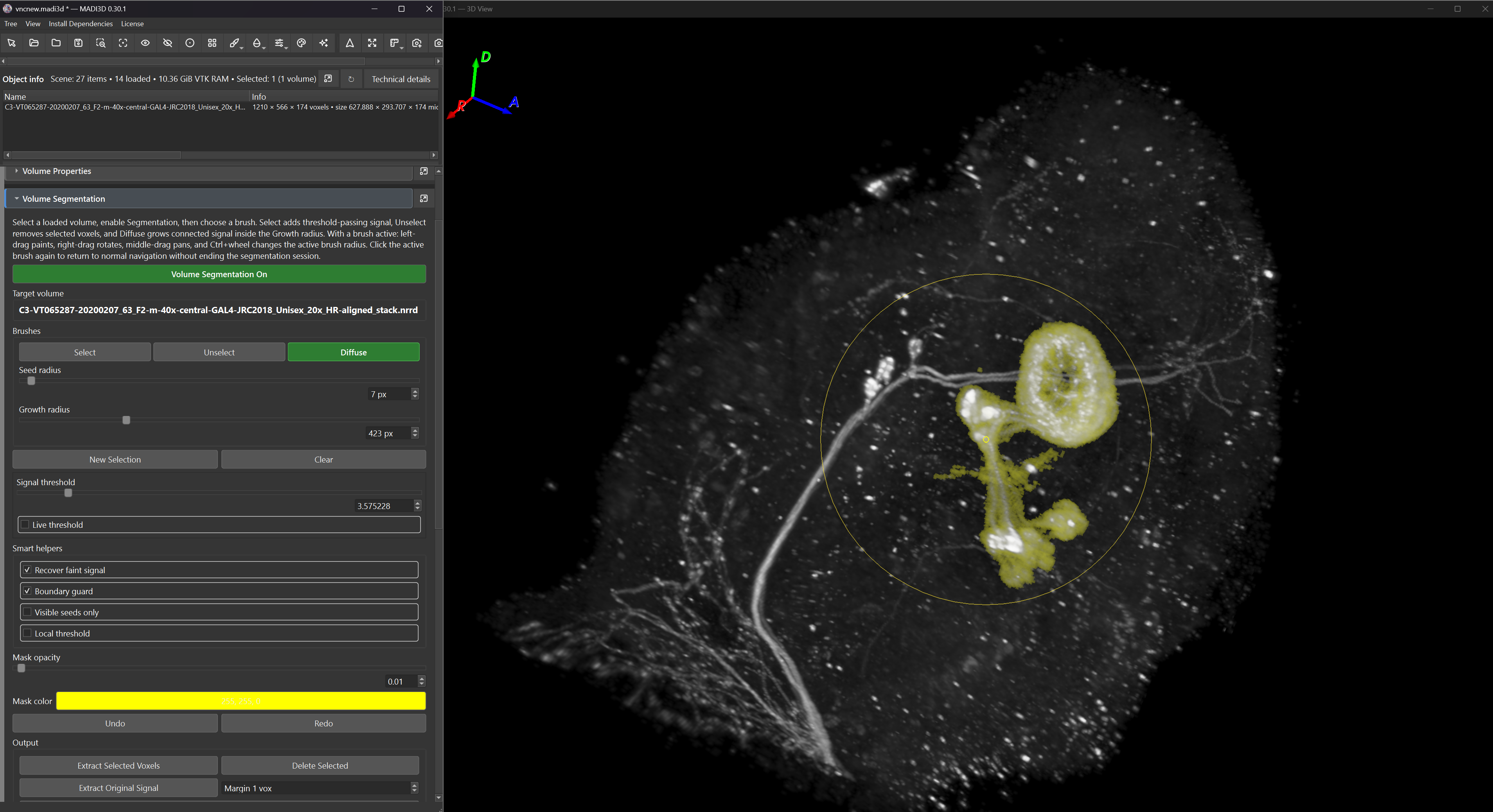
Smart Brush volume segmentation in MADI3D

Volume segmentation

Isolate structures with Smart Brush and extract masks or original signal on the source grid. Longer calculations run in the background with cancellation. Create surface models or prepare segmented signal for neuron matching.

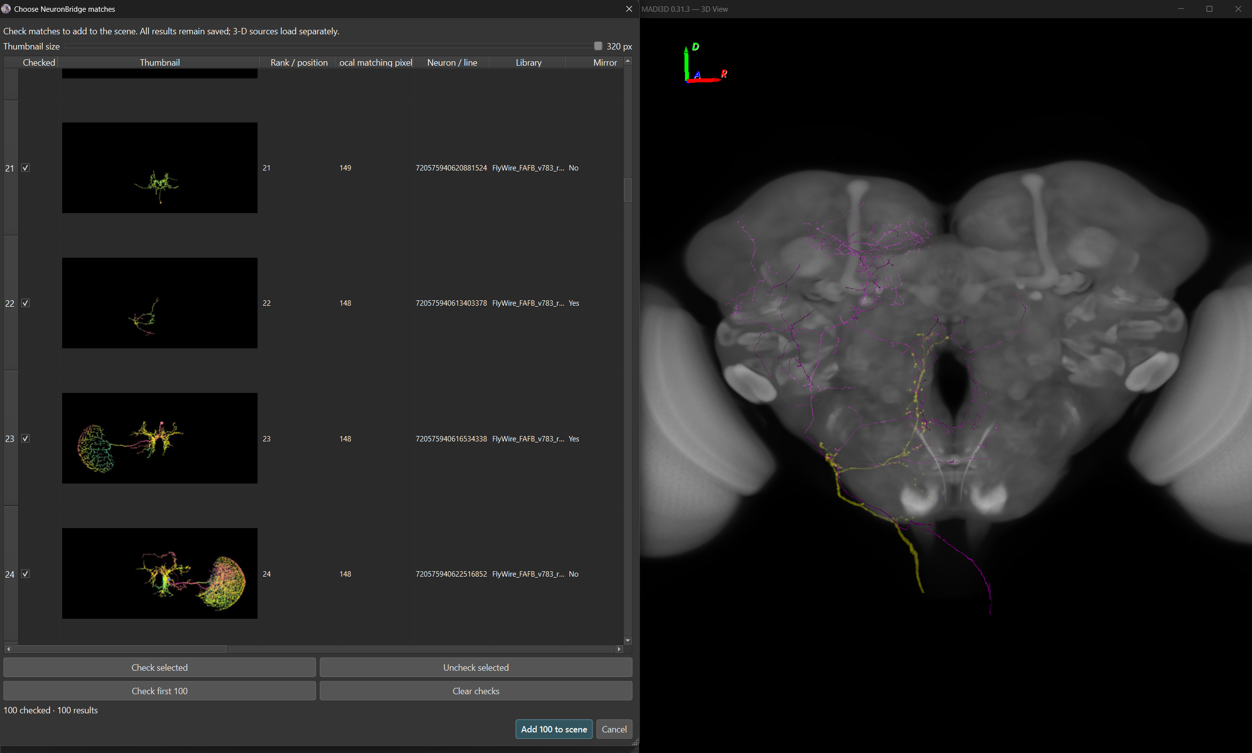
Comparing light and electron microscopy

Move from segmented light microscopy (LM) signal to Color-Depth search and selected electron microscopy (EM) candidates in one workspace. Queries, library snapshots, scores, warnings and result associations remain available when the project is saved and reopened.

Local positive Color-Depth scores are distinct from hosted search scores. Hosted custom-query submission is not included. Morphology search identifies candidates; inspect their full 3D structure before making a biological interpretation. Full-library performance and biological retrieval accuracy have not been established.

Original Fiji ColorMIP Mask Search · Janelia ColorMIPSearch · NeuronBridge · Papers and method references

Example workflow

  1. Import microscopy, select the intended acquisition and channels, and review geometry or calibration warnings. Stitch and review tile placement when needed.
  2. Register microscopy to the appropriate Brain or VNC reference and reformat the neuron channel.
  3. Segment the neuronal signal, preserving its source-grid relationship.
  4. Generate, preview and save a Color-Depth MIP with its alignment evidence.
  5. Install a compatible public library, then search its snapshot locally.
  6. Review the results and load selected candidate LM or EM data.
  7. Compare candidates in 3D and save the project with the query and result evidence.

Continue your investigation in Virtual Fly Brain (VFB): examine neurons of interest alongside annotated anatomy and connectivity, then download available data for further study in MADI3D.

Downloads and help

Download MADI3D

Now available free for Windows x64 from the Microsoft Store. Install from the Store and launch from the Start menu. Store and GitHub updates may become available at different times.

Current public release: MADI3D 0.33.04 Public Beta · 9 October 2026.

Choose the package for your operating system:

GitHub Windows ZIP packages are not code-signed. macOS builds are ad-hoc signed, but not Developer ID signed or notarized. Windows SmartScreen or macOS Gatekeeper may show a first-run warning for these packages.

SHA256SUMS.txt · Release notes and provenance

Qt LGPL source and licenses

Using MADI3D

The public project README provides an introduction and usage notes. For questions or problems, contact the developer or open a GitHub issue.

Scientific references

Find the literature behind the rendering, stitching, registration and neuron comparison methods.

Acknowledgments

With thanks for support and guidance to the University of Cologne, Prof. Dr. Kei Ito and Dr. Thomas Riemensperger.

Support MADI3D

MADI3D remains free to download and use. If you find it useful, voluntary contributions can help cover cloud computing, server costs and automated builds needed to develop and maintain the project.

Contributions go to the developer, not the University of Cologne. No charitable donation receipt is issued.